Cholesteatoma
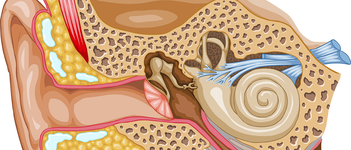
A cholesteatoma is a type of abnormal skin growth that develops behind the eardrum in the middle ear. The growth typically develops as a cyst or sac. Cholesteatomas do not resolve on their own and may increase in size as the sac fills with dead skin cells, fluid, and bacteria. If left untreated, cholesteatomas can affect hearing, balance, and nerve and muscle function in the face. In many cases, they can even destroy the delicate bones of the middle ear.
What Causes Cholesteatomas
In rare cases, babies may be born with the condition. During fetal development, squamous epithelium can become trapped in the middle ear and develop into a cholesteatoma after birth. More often, a cholesteatoma is the result of repeated infections or a poorly functioning eustachian tube. Chronic ear infections or traumatic injury can cause the eardrum to tear or perforate. This allows skin cells to enter the middle ear. If these cells are not shed properly, they can form a cholesteatoma. In cases where the eustachian tube does not allow air to flow through the ear properly to equalize pressure, a vacuum can develop that may cause the eardrum to protrude into the middle ear. This creates a cyst, which collects skin cells, and other debris.
Cholesteatoma Symptoms
Symptoms of a cholesteatoma may initially be mild but increase in severity as the cyst becomes larger. At first, the individual may notice a foul-smelling drainage from the ear or a sensation of plugging and hearing loss. This is typically followed by a feeling of pressure or pain behind the ear and vertigo. If the cyst becomes very large, the patient may experience facial weakness or paralysis and hearing loss. If the cyst becomes infected, it can lead to a life-threatening brain abscess or meningitis.
Diagnosing Cholesteatoma
Most cholesteatomas are diagnosed through a physical examination of the ear using an otoscope or ideally a microscope. The examination will typically reveal a large mass of skin debris deposits in the ear. If the patient is also experiencing dizziness and facial paralysis, the doctor may also order a MRI to rule other potential causes for the symptoms.
Cholesteatoma Treatment
A cholesteatoma typically requires treatment by an otologist / neurotologist. The first step normally involves cleaning the ears followed by ear drops and antibiotics to eliminate any infection. This is typically followed by surgery to remove the cyst. In most cases, the surgery is performed on an outpatient basis using general anesthesia. Some patients experience plugging of the ear following surgery; however, these effects normally resolve within a few weeks. A second surgery may be necessary if the cholesteatoma caused significant damage to the structures or the bones of the inner ear.
Prevention and Long-Term Prognosis
The best way to prevent developing a cholesteatoma is to seek prompt treatment for any ear infections. Anyone experiencing cholesteatoma symptoms should seek treatment as soon as possible to avoid possible complications. The long-term prognosis for cholesteatoma patients is generally good with early treatment. Using modern techniques such as the use of otoendoscopes and lasers in surgery, the chance of recurrence is significantly lowered. Many patients can have their cholesteatomas followed using a special MRI protocol rather than getting a second surgery to check for a recurrence.

